Upload WSI for Analysis

Module 1

Glioma Detection
Uploaded Image

File Name:

Report Preview

Model Result

Based on the model analysis:

Download Report Download

Model Version

M1_v1.0.0-alpha

Category

Model Accuracy

95%

Model Type

Binary Classification

Module Details

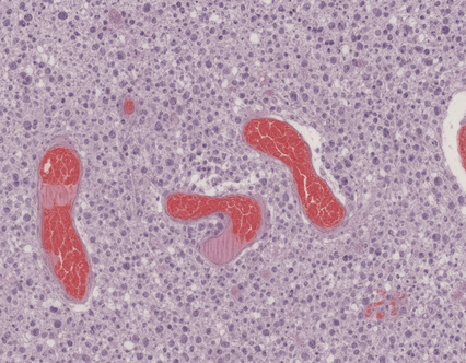
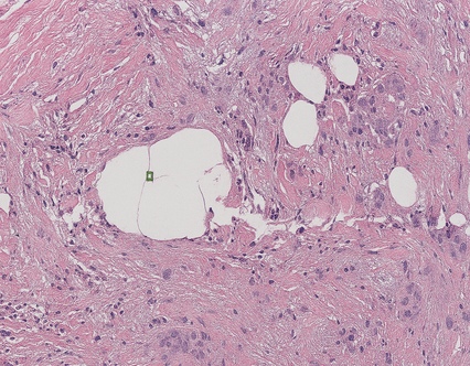
  • Input: Upload a low-resolution whole-slide image (H&E stained) in jpg/png format.
  • Diagnosis Output: Glioma Detected or Glioma Not Detected with a diagnostic report generated from nine quantitative biomarkers.

Interpretation

The following nine quantitative imaging biomarkers provide diagnostic insights based on histopathological feature extraction:

  • Fractal Dimension: Measures complexity and structural irregularity of tumor tissue.
  • Lacunarity: Indicates spatial texture heterogeneity within tumor regions.
  • Entropy: Quantifies cellular randomness and architectural disorder.
  • Short Run Emphasis (SRE): Reflects prevalence of fine-grained textural elements.
  • Long Run Emphasis (LRE): Represents elongated textural patterns, highlighting tumor fiber orientation.
  • Run Percentage (RPC): Assesses texture uniformity, indicative of cellular consistency.
  • Minor Axis Length: Evaluates nuclear size variability, reflecting morphological irregularities.
  • Solidity: Captures compactness and shape regularity of nuclei.
  • Integrated Density: Reflects cumulative staining intensity correlating with cell density.

Predictive Analytics and Disease Prevention

This AI-powered diagnostic module identifies histopathological patterns indicative of glioma, swiftly differentiates glioma-positive from normal tissue. It provides pathologists with rapid and reliable diagnostic clarity, enhancing confidence in clinical decision-making. Part of our comprehensive diagnostic suite, this module is complemented by additional modules offering advanced analytics for detailed tumor subtyping, grading, and further diagnostic insights.

Quantitative Biomarker Analysis

Fractal Dimension

FD

Entropy

EN

Lacunarity

LC

Short Run Emphasis

SRE

Long Run Emphasis

LRE

Run Percentage

RPC

Solidity

SOL

Minor Axis Length

MAL

Integrated Density

ID
AI-Powered Diagnostic Insights from Whole Slide Images

Explore our suite built to accelerate glioma diagnosis*


*Disclaimer: Our solutions assist pathologists by offering rapid, quantitative analyses of histological images. While they enhance workflow efficiency, they are not substitutes for detailed pathological evaluation and should be applied in conjunction with professional expertise.
Module 1
Icon 1
Module 1: Glioma Detection

Rapid, AI-driven histopathology analysis to detect or exclude glioma from tissue samples.

  • Input: Upload a low-resolution WSI (H&E-stained) in jpg/png format.
  • Output: Glioma Detected or Glioma Not Detected.
  • Accuracy: 95% on internal validation.

This module supports early-stage glioma identification by analyzing histopathological patterns, providing reliable diagnostic clarity. It forms the foundation for subsequent modules focused on detailed subtyping and grading.
Module 2
Icon 2
Module 2: Glioma Subtype Classification

AI-driven histopathology analysis to classify glioma subtypes.

  • Input: Upload a low-resolution WSI (H&E-stained) in jpg/png format.
  • Output: Normal, or one of the three glioma subtypes: Astrocytoma, Oligodendroglioma, or Glioblastoma Multiforme (GBM).
  • Accuracy: 90% on internal validation.

This module enables glioma subtype identification from images, empowering early clinical insights essential for patient-specific treatment planning. It paves the way for advanced whole-slide analysis in the next module, supporting deeper pathological interpretation from the model.
Module 3 WSI-Based Glioma Subtype Diagnosis
Diagnosis Icon
Module 3: WSI-Based Glioma Subtype Diagnosis

AI-driven whole-slide histopathology to identify and highlight key diagnostic regions.

  • Input: Upload a high-resolution WSI (H&E-stained) in svs format.
  • Output: Glioma subtype classification with visual interpretation graph and key region descriptors.
  • Accuracy: 95% on internal validation.

This module supports glioma subtype diagnosis from WSIs by identifying diagnostically relevant regions, providing visual interpretations, and generating clear, region-specific morphological descriptions. It enables pathologists to efficiently review key areas and make confident clinical decisions.

Hi There


Start a Conversation


ViviAI Team here ....

Hi There


The team typically replies in few minutes.

ViviAI Bot

ViviAI